Glaucoma is a disease that gradually decreases visual field and often occurs after the age of 40.

“What symptoms should I see an ophthalmologist for?”
“Is it true that glaucoma can lead to blindness as it progresses?”
“Is there a good way to prevent or treat glaucoma?”

Answers to the above questions will be explained in this article, as well as what glaucoma is, and its causes and symptoms.

  • what kind of disease is glaucoma
  • About symptoms and causes
  • How to prevent and treat glaucoma

Glaucoma is a common disease for middle-aged and older adults, but in the early stages there are almost no subjective symptoms. However, it is also possible to stop or slow the progression of the glaucoma if there is early treatment. For early detection, it is helpful to schedule regular eye examinations.

What is glaucoma?

Light entering the human eye forms an image on the retina at the back of the eyeball. Only when electrical signals are transmitted from the retina to the brain is “seeing”. Glaucoma is a disease that damages the optic nerve (retinal nerve fibers) that transmits visual information.

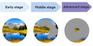
It is a disease in which the bundles of nerve fibers, originally said to number 1.2 million, are reduced, and as the optic nerve is reduced, the field of vision gradually narrows. It progresses slowly until the vision is affected, and there are almost no symptoms in the early stages.

If left untreated, glaucoma can lead to loss of vision and eventually blindness. More than 10% of all people over the age of 60 suffer from glaucoma, making it the number one cause of blindness in Japan. At the age of 40 or older, see an ophthalmologist.

Once glaucoma is diagnosed, it is important to visit the hospital regularly for treatment to control and slow the progression of the disease as much as possible.

Intraocular pressure deeply associated with glaucoma

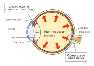
The optic nerve damage occurs when intraocular pressure (pressure inside of the eye) increases. Normal intraocular pressure is generally 10~21mmHg , but the intraocular pressure that the optic nerve can withstand (healthy  intraocular pressure) varies from person to person. Therefore, it is believed that optic nerve damage can be controlled by reducing intraocular pressure that has risen above a tolerable level.

If the intraocular pressure becomes abnormal, the optic nerve will naturally be damaged and, despite the normal values, some people may experience visual impairment. It should be noted that “what level of intraocular pressure is safe” varies from person to person.

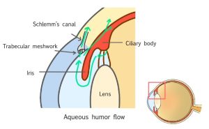
The fluid in the eye called aqueous humor affects intraocular pressure. The role of the aqueous humor is to carry various nutrients inside the eye instead of blood. The aqueous humor, which is produced in the ciliary body behind the iris, is discharged from the angle  between the iris and the cornea through a filter-like tissue called the trabecular meshwork and out of the eye through a tube called Schlemm’s canal.

Intraocular pressure depends on the amount of aqueous humor and the resistance of the aqueous humor to drain. The flow of aqueous humor will stop and/or the outlet will clog, increasing intraocular pressure.


Types of glaucoma

There are several types of glaucoma. Causes, symptoms, and people prone to it vary.

Angle-closure glaucoma

The outlet of the aqueous humor is called the angle. Angle-closure glaucoma is caused by an increase in intraocular pressure as a result of narrowing of the angle, which prevents the flow of aqueous humor.

  • Acute type: Significant increase in intraocular pressure, accompanied by decreased visual acuity, eye pain, headache, and nausea
  • Chronic type: may be accompanied by blurred vision especially at night or mild headaches

Women in their 40’s and older are 3-4 times more prone to the disease than men, especially those with farsightedness.
There are certain drugs that cannot be used for those diagnosed with angle closure glaucoma. Must check with your ophthalmologist regarding your glaucoma type and the drug’s should not be used or not.

Open-angle glaucoma

Even if the angle of the anterior chamber is wide enough, they may become clogged with trabecular meshwork. As a result, the increased intraocular pressure causes open-angle glaucoma.
Intraocular pressure rises slowly, with almost no symptoms in the early stages. As the disease progresses, some areas of difficulty in seeing may appear and visual field defects may begin to occur.
Many diseases occur over the age of 40, and the rate increases with the age. It is said that it tends to be more common in people who are myopic originally. POAG occurs more frequently in eyes with myopia.

Normal tension glaucoma

In some cases, glaucoma develops even when the intraocular pressure is within the normal range of 10 to 21mmHg and is classified as part of open-angle glaucoma. It is caused by a weak structure of the optic nerve papilla, which cannot withstand even normal intraocular pressure and damages the optic nerve.
A previous study of several thousand people reported that glaucoma occurred in 5% of the population aged 40 years or older, and that more than 70% of them had normal tension glaucoma in Japan.

Secondary glaucoma

Secondary glaucoma is caused by the effects of another disease that was originally present. It may be caused by due to prolonged steroid medication eye drops, retinal diseases led by diabetes or etc.
Prolonged use of steroid application, oral medications, or eye drops can increase intraocular pressure, therefore requires an ophthalmologist’s visit.
Secondary glaucoma also includes increased intraocular pressure caused by trauma, corneal disease, or inflammation of the eye, which results in glaucoma.

Childhood (congenital glaucoma)

If the child is born with developmental abnormalities of angle, the intraocular pressure increases, and the optic nerve is damaged.
Occurs in about 1 in 30,000 people, the early-onset type occurs in approximately 80% of patients by the age of 1, while the late-onset type occurs in younger people in their teens to 20s, although the degree of the angle abnormality is less severe.

Glaucoma symptoms

Early glaucoma has few warning signs or obvious symptoms and tends to be detected late.
Open-angle glaucoma, for example, does not manifest itself until it is quite advanced, and patients themselves rarely notice that they are losing their vision.
Similarly, angle-closure glaucoma has no symptoms when the intraocular pressure is normal in many cases, but the situation changes when an attack occurs. Especially in the case of acute attacks, the pressure jumps to a level of 40 to 60mmHg, resulting in reduced visual acuity and significant headaches and eyeaches..

(Images of the loss of visual field due to the progression of glaucoma)

Exams for diagnosing glaucoma

Glaucoma, which has few subjective symptoms, is difficult to self-check in its early stages. Regular eye doctor visits and early detection are the best ways to reduce the progress of glaucoma.

Intraocular pressure Exam

Intraocular pressure exam includes measuring methods by using air-puff on the eyeball or by compressing the cornea after eye anesthesia is administered.
However, it is difficult to correctly determine intraocular pressure with a single measurement, as the value tends to fluctuate during the day and tends to be higher in winter.
Since lowering intraocular pressure is the basis of glaucoma treatment, measurement of intraocular pressure is an important exam.

The angle exam

The condition of the angle is examined by shining a light on it with an exam-contact-lens placed over the eye for testing.
Since the examination is performed under ophthalmic anesthesia, pressure is felt but no pain is felt.
Narrow angle indicates that the patient is a type that is prone to acute attacks of glaucoma.

Fundus exam

Funduscopy involves observing the back of the eye with the pupil dilated to determine the degree of optic nerve damage.
The shape, depth, size and hemorrhage of the optic nerve head and the status of optic nerve defects can be confirmed.
In recent years, the equipment “Optical Coherence Tomography (OCT)” has become popular. Being able to measure the thickness of nerve fiber layer, etc. in the image, we expect early detection of glaucoma because easy to notice abnormalities of early stage.

Visual field exam

Tests how light appears in the visual field without moving the eye. The lack of vision can be checked by pressing a button when a light is seen on the testing machine.
This method is also used to periodically check for progressive visual field loss and to determine the level of glaucoma.

How to treat glaucoma

The most important part of glaucoma treatment is to lower intraocular pressure so that the optic nerve is not reduced.
Although there are individual differences, the optic nerve is stretched like a cluster of about 1.2 million fibers. However, damaged optic nerves do not recover. There is also glaucoma for which lowering the intraocular pressure does not slow the rate of progression. Therefore, it is important to know that the goal of treatment is to slow the progression of the disease as much as possible, not to reverse it.

Medication

Eye drops are first prescribed to treat glaucoma.
Broadly speaking, eyedrops have two main effects.

1.Promotes drainage of the aqueous humor
2.Suppresses the production of aqueous humor

Additionally, there are drugs that are intended for both of these purposes.
Usually, you would start with one type of eye drop and if the effect is weak, additional types of eye drops would be prescribed.
One drop should be applied at a time, with an interval of at least 5 minutes when multiple medications are used together. This is believed to be effective in maximizing efficacy while minimizing side effects of the drug.

Laser treatment

There are two types of laser treatment for glaucoma.

Iridotomy Punctures the iris to improve the flow of the aqueous humor. Effective for angle closure glaucoma
Trabeculoplasty Laser irradiation of trabecular meshwork to promote aqueous humor drainage Effective for open-angle glaucoma


The iris is the coloured circular part of that eye that surrounds the black pupil (= central part). On the other hand, the trabecular meshwork is located where the cornea meets the iris.
Both of these procedures use ophthalmic anesthesia, so there is little or no pain. It is also available on an outpatient basis.


Surgery

If continued eye drops and laser therapy are not sufficient to control glaucoma, surgery is considered.

1.Trabeculotomy, in which the trabecular meshwork is incised to facilitate the drainage of aqueous humor
2.Filtration surgery to allow the aqueous humor to seep out of the eye

However, surgery is also performed not to improve symptoms, but only to lower intraocular pressure and slow the progression of the disease. Other surgeries include connecting tubes in and around the eye to guide aqueous humor and lower intraocular pressure.

How to prevent glaucoma

Once damaged, the optic nerve does not recover, so early detection and initiation of treatment is important to prevent glaucoma from worsening and to maintain visual field. However, it is difficult to notice glaucoma by yourself because there are few subjective symptoms in the early stages. It is a good idea to have an eye exam regularly after the age of 40.
While glaucoma cannot be completely prevented, here are a number of ways to get risk factors away:

Exercise to improve diabetes, sleep apnea syndrome, etc.

People with the following diseases and symptoms are said to be more prone to glaucoma.

  • Diabetes
  • Migraines
  • High myopia and hyperopia

It is a good idea to make a conscious effort to engage in light jogging or walking. Proper exercise can also lead to increased blood flow in the eye.

No smoking to ensure capillary feeding

The harm of tobacco as it relates to glaucoma is the effect of nicotine and carbon monoxide on blood vessels. Hemoglobin function is reduced, and blood circulation is impaired, which tends to result in a lack of nutrients and oxygen to the inside of the eye.
We do not recommend smoking as it can accelerate the development and progression of glaucoma.

Avoid intraocular pressure build-up

Pressing the eyelid in or closing the eyelid tightly temporarily increases intraocular pressure. Tightening the neck also leads to increased intraocular pressure.
When sleeping, a supine or sideways position with the glaucomatous eye down is also considered undesirable.

Living the right way

There is nothing that is forbidden in everyday life after receiving a diagnosis of glaucoma. Drinking alcohol in moderation is not a problem, and it is also a good idea to take up hobbies such as travel and sports.

Q&A about Glaucoma

Q: What if I am suspected of having glaucoma during a health check?

You should have been advised to see an ophthalmologist. Since there are virtually no subjective symptoms in the early stages of glaucoma, patients should be examined promptly by an ophthalmologist before any abnormalities are felt in the eye.

Q: What should a person with glaucoma not do?

No special requirements. You can use your eyes for exercise, reading, etc. However, remember to use eye drops on a daily basis. Please see an ophthalmologist regularly.


[Editorial Supervision]

Professor Takashi Fujikado

Specializes in pediatric ophthalmology, amblyopia strabismus, ophthalmology and low vision, as well as general ophthalmology.

This browser will not be covered by the display operation guarantee.
To view the HOYA site, please switch to a supported browser.
After 2022, the browsers recommended for viewing and operation on Windows 10 are as follows.

Please contact the browser provider for download and installation methods.

×