The retina is the most important part of vision, as it detects light and color. Retinal detachment occurs when retinal tissue separates and, if left untreated, can lead to vision loss or blindness.
Therefore, it is important to detect retinal detachment as early as possible and begin treatment.

This article describes the following topics.

・Causes of retinal detachment include eye damage, disease, and aging
・Symptoms of retinal detachment include floaters and photopsia
・Treatment varies depending on the stage

What is Retinal Detachment

The retina receives light that enters the eye as a stimulus and transmits it to the brain. The retina is composed of 10 layers of tissue, the deepest of which is called the retinal pigment epithelium. Retinal detachment is a disease in which the retina separates from the retinal pigment epithelium, resulting in impaired vision.

Retinal detachment itself is painless, so it is difficult to notice on your own, but floaters may appear as a warning sign. (For more information on floaters, see this article.)

The macula, located in the center of the retina, is sensitive to light and is an important part of vision. If retinal detachment progresses to the macula, vision will rapidly decrease. If left untreated, it can lead to blindness, and vision may not be restored even with treatment.

There are several types of retinal detachment. The most common type is called rhegmatogenous retinal detachment, which is caused by a retinal break, such as a small hole or tear. Non-rhegmatogenous retinal detachment occurs when diabetic retinopathy progresses, while tractional retinal detachment is typically caused by fibrous proliferation after vitreous hemorrhage.

Causes of Retinal Detachment

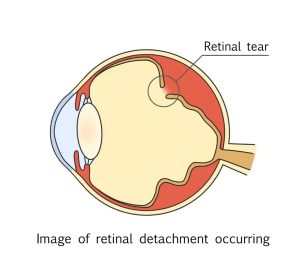
A typical retinal detachment begins with a retinal tear. If left untreated, the tiny hole allows vitreous fluid to penetrate between the retina and the layers underneath, eventually causing a retinal detachment.
Rhegmatogenous retinal detachment caused by a hole is most common in 20s and over 50s and older. People with severe myopia or a family history of retinal detachment are more likely to develop retinal detachment.

Age-Related Posterior Vitreous Detachment

The vitreous body inside the eye liquefies with age. When the liquefied vitreous separates from the retina, it can pull the retina apart, causing a hole or detachment of the vitreous and retina together.
Approximately half of people in their 60s and more than 70% of people in their 70s develop vitreous detachment, but in the case of myopia, it is said to occur about 10 years earlier.

Tear due to bruising or trauma

Some people may think of boxing when they hear the term “retinal detachment.” This is because a certain number of boxers suffer from retinal detachment due to the impact of receiving a punch. The impact of being hit by a baseball or soccer ball can also damage the retina.

High myopia

Myopia is often caused by an elongated eyeball. The longer the eyeball, the thinner the retina becomes, making it more susceptible to retinal detachment.

Atopic dermatitis and other diseases

Retinal detachment is also likely to occur in people with severe skin inflammation around the eyes, such as atopic dermatitis. It is thought that repeated rubbing of the eyelids due to itching puts pressure or impact on the retina, causing retinal detachment.
Other conditions, such as diabetes, may also cause this condition.

Symptoms of Retinal Detachment

Typical symptoms of retinal detachment include the following. If you have any of these symptoms, it is best to have an examination as soon as possible.

Floaters – looks like small insects or debris-like objects appear to fly in front of the eye.
Photopsia – Flickering at the edges of your visual field. Lightning-like flashes in the dark.
Visual field defect – Objects appear distorted or your field of vision becomes narrower and you cannot see parts of them.
Deterioration of vision – makes it difficult to see clearly what you want to see.

How to Test for Retinal Detachment

If the exam reveals a retinal detachment, treatment should be sought as soon as possible. Early detection and prompt treatment are important to minimize the deterioration of vision.

Fundus examination

Thirty minutes before the examination, a dilating eye drop will be applied to dilate the pupils and a fundus examination will be performed. The examination will reveal the extent and range of retinal detachment or retinal break, as well as the condition of the vitreous body.
The test is painless, but the eye drops will blur your vision and prevent you from driving for a few hours.

Visual field testing

The visual field test is performed by using a testing machine and pressing a button when a light is seen to determine if a visual field defect has occurred. In everyday life we use both eyes, we often do not notice if part of one eye’s vision is missing.
That’s why, even if you notice a slight change, it’s important to see an eye doctor as soon as possible before the condition progresses.
In addition, visual acuity tests and OCT (optical coherence tomography) tests are also performed to determine if the retinal detachment has extended to the macula.

If you are diagnosed with physiological floaters

If a patient is concerned about floaters and visits an ophthalmologist but there are no retinal tears, these could be age-related changes in the vitreous without a retinal detachment, there is no need to worry right away. However, if the posterior vitreous detachment progresses with age, there is a possibility of retinal detachment. Therefore, it is advisable to confirm with an ophthalmologist when and how to follow up.
If new symptoms are added afterwards, such as worsening of flying mosquitoes or seeing light (photopsia), do not wait for the follow-up period, instead it is best to consult the ophthalmologist as soon as possible.

Treatment of Retinal Detachment

The treatment strategy varies depending on the condition of the retina and the progression of the retinal detachment.

1.Retinal tear but no retinal detachment

If a retinal tear has occurred but the retina has not detached, a laser may be used to coagulate the retina around the tear and prevent the detachment from progressing.
The retinal pigment epithelium, the lowest layer of 10 retinal layers prevents vitreous fluid from flowing into the upper layer (called the neural retina) and prevent the retina separating from the retinal pigment epithelium. However, depending on the size of the tear and the pulling force of the vitreous body, the preventive effect of treatment varies.

2.When the retina has begun to detach

If retinal detachment has already begun, surgery is required to fix the retina in place.
Surgery is performed in two main ways.

The first is called retinopexy, in which a sponge-like object is placed over the area of the retinal tear from outside the eye and then using thermal- or cryo-coagulation the detached retina around the break is re-attached. If necessary, fluid that has accumulated under the retina may be drained.


The other, called a vitrectomy, involves inserting small surgical tools into the eye to treat the retinal detachment from inside the eye. Air, a special gas, or silicone is injected to hold the detached retina in place, and the vitreous is removed.


Q&A about Retinal Detachment

What causes retinal detachment?

Retinal detachment can be caused by aging, diseases such as diabetic retinopathy, or trauma such as a blow to the eye. It begins as a tear or hole in the retina and eventually progresses to retinal detachment.

What are the signs of retinal detachment?

The first sign is often the appearance of flying mosquitoes. There is no pain during retinal detachment, but the irritation caused by the detachment can be transmitted to the brain, causing a sudden flash of light (photopsia).

How long does it take for retinal detachment to progress to blindness?

The rate of progression varies from person to person and is also dependent on the progression of the disease, so it is difficult to generalize, but elderly patients often have liquefied vitreous and tend to have more rapid progression of retinal detachment, and visual field loss may spread within a week. In such cases, the patient must rest and undergo surgery immediately.

[Editorial Supervision]

Professor Takashi Fujikado

Specializes in pediatric ophthalmology, amblyopia strabismus, ophthalmology and low vision, as well as general ophthalmology.

This browser will not be covered by the display operation guarantee.
To view the HOYA site, please switch to a supported browser.
After 2022, the browsers recommended for viewing and operation on Windows 10 are as follows.

Please contact the browser provider for download and installation methods.

×