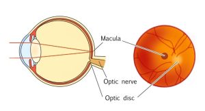
The macula, where photoreceptor cells that distinguish shapes and colors are concentrated, is an important tissue that greatly affects visual acuity. However, with age, bleeding and swelling of the macula may occur and visual acuity is reduced. This is an age-related macular degeneration (AMD), a condition that can lead to blindness if left untreated.

“Can anyone notice the sign of AMD?”
”How do I check it out myself?“
“How should I treat it?”

Such questions shall be explained in this article, as well as:

・What is AMD?
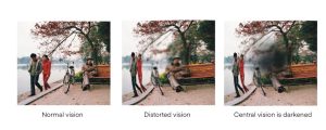
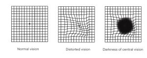
・How does the onset of the disease change the way you see?
・How to prevent AMD

Since this disease can occur to anyone over the age of 40, it is important to see an ophthalmologist regularly even if there is nothing wrong with your eyes. Early detection and early treatment are key to maintaining healthy vision.

What is Age-related macular degeneration(AMD)

The macula is located in the center of the retina, where the photoreceptor cells that distinguish shape and color are densely clustered. Although the macula is a small area of the entire retina, it is the main area used to see things; consequently, abnormalities in the macula can cause a significant loss of visual acuity.
Age-related macular degeneration (AMD), is a disease that causes bleeding and swelling in the macula, resulting in vision loss.

The main cause is age-related.

・Object appears distorted
・Darker center of vision
・Lack of center of vision
・Reduced visual acuity

The above symptoms often appear.

If left untreated, AMD will progress gradually (or rapidly), and in advanced cases, there is no hope of restoring vision through treatment. For this reason, along with diabetic retinopathy and glaucoma, it is feared that in the worst-case scenario, the disease can lead to blindness.
Although it has long been known as a major cause of blindness in adults in Europe and the United States, the number of patients with AMD is increasing in Japan due to dietary changes and aging populations.

Types of AMD

There are two types of AMD: Wet AMD and Dry AMD.
The outer layer of the retina is called the retinal pigment epithelium (orange in the illustration), outer of which, there is the choroid lined with blood vessels that deliver nutrients to the retina.

Wet AMD

In the Wet AMD, “neovascular vessels” from the choroid extend and reach above the retinal pigment epithelium, damaging the retina, including the macula. Newly formed blood vessels are very fragile, and water serous component in the blood can leak out or the vessels can break, causing bleeding inside the retina. This results in poor vision and acute loss of vision.
This type of SMD requires early exam and treatment and is common among Japanese.

Dry AMD

This is a disease in which waste accumulates inside the retina as   it ages, and the macula to atrophy along with the retina itself. The progression is slow, but it may change to Wet AMD in the middle of the process, resulting in acute vision loss.

Risk factors of AMD

The main cause of AMD is age-related damage to the macula, which plays an important part in vision.
In addition to aging, the following factors are also indicated:

・Smoking
・High blood pressure
・A diet high in saturated fat
・Obesity
・Ultraviolet light
・Genetic, etc.

It has been estimated from these factors that AMD has increased significantly in recent years. A survey of residents of the town of Hisayama in Fukuoka Prefecture in Japan found that AMD had increased to 1 in 80 (1.3%) in 2007 from less than 1 in 100 people over age 50 (0.87%) in 1998.
When the increase in the elderly population is taken into account, the estimated number of patients has almost doubled from an estimated 370,000 to 690,000 in Japan over the past nine years. It is expected to increase further as the population ages.

The symptoms of AMD

When macular degeneration occurs, the following symptoms usually appear:

Distorted vision

In the early stages of AMD, the symptoms of heterophoria, metamorphopsia in which objects appear distorted, may appear. This is caused by a disruption in the arrangement of photoreceptor cells. The appearance of the peripheral areas is not distorted.
Sometimes, only centered objects may be difficult to see, and the center of the vision may be blurred, or hazy.

Center vision is darkened

As AMD progresses, the center of vision becomes darker and progressively less visible. This is called central scotoma and is a characteristic condition of AMD.
Gradual loss of color discrimination can also occur.

Reduced visual acuity

More symptoms may lead to reduced visual acuity, and some people may experience reduced visual acuity at a rapid pace. In most cases, the eye does not feel pain, but if left untreated, it can lead to blindness.

AMD exam sequence

Early and regular eye exams are important for early detection and treatment of AMD.
At the time of the visit, the following exams will be performed.

Medical interview

First, you will be asked what subjective symptoms you have and how long you have felt them, so answer as accurately as possible. In addition, you may be asked about your smoking habits, non-ocular illnesses, and medications.

Vision screening

Since AMD may be the cause of vision loss, the first step is to examine vision. This is particularly important when you feel your vision fades rapidly over a short period of time.

Amsler grid testing

The Amsler grid test is an essential test for the diagnosis of AMD. In this test, the center of the grid is viewed with only one eye, and if there are symptoms of AMD, the following abnormalities may appear.

・center appears distorted
・missing the center
・looks black

Optical coherence tomography (OCT scanning)

Cross-sectional examination of the macula using an instrument called an optical coherence tomograph provides a three-dimensional view of retinal swelling and choroidal neovascularization.
Because it does not use contrast dye, it is characterized by its low burden on the patient’s body and is often performed continuously during follow-up.

Fundus exam

This is an examination that has a main focus of determining the state of bleeding in the macula. Eye drops are used to open the pupil for a closer look at the retina.

Fluorescein angiography

This is done when you examine the neovascular structures in more detail. The test involves injecting a fluorescent dye into a vein in the arm and then photographing the fundus to determine the location, shape, and activity of neovascular vessels.

AMD treatment

Even if you develop AMD, you may be able to maintain or improve your vision, depending on the timing and method of treatment. It is advisable to attend regular eye exams to monitor the progress of the disease, as well as to review one’s diet and exercise moderately. Lifestyle modifications such as UV protection and smoking cessation may also be effective.
In Wet AMD, the following treatments are used (There is currently no effective treatment for Dry AMD.)

Anti-VEGF therapy

Proteins in blood vessels, called ” VEGF (Vascular endothelial growth factor)”, are said to grow neovascular and damage macula. Therefore, when treating AMD, this anti-VEGF drug is injected into the eye to inhibit neovascular activity.
It is minimally painful since it is injected through local anesthesia. Anti-VEGF therapy can also be administered outpatient setting, but three days before and a few days after injection, anti-microbial instillation eye drops are needed to clean the eye.
Usually, two to three injections are given at intervals of 1 to 1.5 months, followed by another injection if the neovascularization recurs, for continuous treatment.

Photodynamic therapy

This method involves administering a drug that reacts to a specific light (laser) and applying a laser when the drug reaches the neovascular vessels. Drugs activated by the laser occlude the vessels, preventing fluid in the blood from leaking out.
Often used in combination with anti-VEGF therapy, it is good in terms of that it has virtually no affection on anything other than neovascular vessels. The examination is performed every few months and the treatment is continued if the patient needs to be treated again.

Laser Photocoagulation

The procedure uses a laser to burn and close the neovascular vessels, but may also damage the surrounding normal tissue.
When laser photocoagulation is used, the neovascular vessels must be located away from the macula for safety.

How to prevent AMD

Many people who notice AMD and visit an ophthalmologist find that the disease has already progressed considerably. If you notice any change in vision, see an ophthalmologist as soon as possible.
Fundus exam performed during a health check may reveal waste products to the retina. This is one of the signs of AMD. Continued follow-up with regular visits afterwards will help prevent the onset and worsening of symptoms.

Quit Smoking

Studies show that smokers are at high risk of AMD. The probability of developing macular degeneration after 5 years is 5.2% for more than 20 cigarettes a day, and the risk of smokers is also referred to as 1.7 to 3.3 times greater than non-smokers.
Quitting smoking is essential for the prevention of AMD.

UV rays protection

Ultraviolet rays are mostly absorbed by the cornea, but when some of them reach the retina, the retina is damaged. To counteract this, it is recommended to wear sunglasses with UV protection and a hat.
Blue light emitted from computers and televisions is also considered to be bad for the eyes, so care should be taken to avoid prolonged use.

Consumption of green and yellow vegetables

Green and yellow vegetables often contain lutein and zeaxanthin present in the macula pigment.
Therefore, foods containing vitamin E, vitamin C, beta carotene, etc., known as antioxidant vitamins, as well as minerals that make up antioxidant enzymes, should be consumed.

・Containing vitamin E, Vitamin C, Beta Carotene and more…carrot, pumpkin, soya, brown rice, etc.
・Containing minerals (e.g. zinc) … Seaweed, Oyster, etc.

When it comes to eating, an overall balance of nutrition is important. To avoid obesity, it is important to incorporate moderate exercise.

Q&A about AMD

Q: Who is prone to AMD?

AMD is a condition that can occur to anyone over the age of 40. Older men in particular are at increased risk of developing the disease. Smokers are also more likely to develop AMD than non-smokers.

Q: Is AMD a curative disease?

Once developed, AMD is difficult to cure completely, and the patient is expected to live with it for a long time to minimize its progression as much as possible.
With proper treatment, symptoms can be somewhat improved or maintained. In some cases, delayed treatment can lead to vision loss, so be sure to see an ophthalmologist immediately if you have any symptoms that concern you.

Q: How long does it take to cause blindness after AMD?

If left untreated, AMD can lead to blindness. However, it is impossible to say how long it will take to reach blindness. AMD can be Wet AMD, which causes rapid vision loss, or Dry AMD, which progresses slowly, and the time until vision loss varies from person to person.

[Editorial Supervision]

Professor Takashi Fujikado

Specializes in pediatric ophthalmology, amblyopia strabismus, ophthalmology and low vision, as well as general ophthalmology.

This browser will not be covered by the display operation guarantee.
To view the HOYA site, please switch to a supported browser.
After 2022, the browsers recommended for viewing and operation on Windows 10 are as follows.

Please contact the browser provider for download and installation methods.

×