Everyone has difficulty seeing in the dark, but if your vision seems to deteriorate more rapidly at night or in dark places than in bright places, you may have night blindness. Some types of night blindness cannot be treated and may lead to vision loss.

This article describes following topics.

・Characteristics and Causes of Night Blindness
・Methods of examination
・How to treat and cope with it

It is important to see an ophthalmologist as soon as possible and to continuously check the progress of the disease.

What is Night Blindness?

Night blindness is a condition that makes it difficult to see in the dark or at night.

The retina has photoreceptor cells that work in bright light (cone cells) and photoreceptor cells that work in dim light (rod cells). People with night blindness have difficulty seeing in dim light because the rod cells that receive information when it is dark do not function normally.

What is dark adaptation?

If a room suddenly becomes pitch black, you won’t be able to see anything for a while, but over time your eyes will get used to the darkness. This reaction to adapting to the dark is called “dark adaptation.”

Normally, it takes about 30 minutes for dark adaptation to begin and end. However, if it takes 2 to 3 hours to “get used to the darkness,” dark adaptation is likely delayed.

People with night blindness cannot adjust their eyes to darkness because they cannot see.

Congenital and Acquired Night Blindness

There are two types of night blindness: congenital night blindness, which is caused at birth, and acquired night blindness, which is caused by nutritional deficiencies or inflammation.

Characteristics of Congenital Night Blindness

Congenital night blindness is further classified into two types: congenital night blindness that develops during childhood and progresses gradually, and non-progressive night blindness, in which the symptoms do not progress even after onset.
A typical example of the progressive type is retinitis pigmentosa, which is designated as an incurable disease in Japan and whose symptoms often appear by the age of 20 and progress over several decades. In addition to night blindness, symptoms such as narrowing of the field of vision and decreased vision may also appear. Symptoms such as glare sensitivity also appear, and ultimately lead to blindness.

For more information, please see the article “Retinitis Pigmentosa.”

On the other hand, non-progressive night blindness is known to be caused by hereditary diseases such as Oguchi disease and fundus albicans. In most cases, there are no abnormalities in the visual field, visual acuity, or color vision, but the patient’s dark adaptation is slow, making it difficult to walk in the dark.

Characteristics of Acquired Night Blindness

Acquired night blindness may not become evident until adulthood. Causes include vitamin A deficiency, cancerous retinopathy, and ocular iron rust. Night blindness may also occur as a result of advanced glaucoma, diabetic retinopathy, and other conditions.
Vitamin A deficiency used to be caused by malnutrition, but now it is mostly caused by digestive disorders or liver damage.

Examination and Diagnosis of Night Blindness

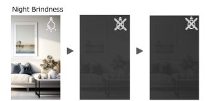
If symptoms of night blindness are present, the ophthalmologist will perform the following tests

Fundus examination

This is an examination to check the condition of the blood vessels in the fundus, the retina, and the optic nerve. In the case of retinitis pigmentosa, pigmentation and uneven deposition can be seen in the retina, but in the early stages or in mild cases, it may not be possible to confirm this, so observation is necessary.

Visual field test

This is a test to determine the extent of the visual field, or how wide the patient can see. Since retinitis pigmentosa and acquired night blindness tend to cause narrowing of the visual field, the test can be used to determine the stage of the disease.

Electroretinogram (ERG: electroretinogram)

This is a test to determine whether the retina is working properly. It examines the function of the retina by observing the electrical changes that occur when the retina is exposed to strong light.
If the function of rod cells, which function in the dark, is impaired, evoked potential becomes smaller or no response is observed.

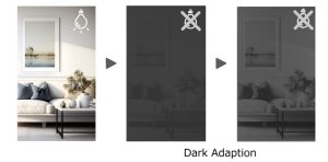
Dark adaptation test

The dark adaptation test is used to determine the degree of night blindness. This test measures how the eye adapts to light when a subject moved from a brightly lit place to a dark room.
A dark adaptometer can be used to construct a dark adaptation curve and check the response of both cones and rods, the absence of rod response being characteristic of night blindness.

Optical coherence tomography (OCT)

OCT is a test that examines a cross section of the retina. If retinitis pigmentosa is advanced, this test can show areas of photoreceptor cell loss.

Fundus autofluorescence (FAF: fundus autofluorescence)

Autofluorescence is the emission of longer wavelength fluorescence after exposure to light of a specific frequency. The image is bright when there is a large amount of lipofuscin, a pigment present in the retinal pigment epithelium, and dark when the lipofuscin decreases as the disease progresses and the retinal pigment epithelium is damaged.
Regular check-ups are recommended to monitor the progression of the disease.
Other tests that may be performed if acquired night blindness is suspected include blood vitamin A levels, the presence of tumors, and the presence of autoantibodies against the retina.

Treatment of Night Blindness

There is no established treatment for congenital night blindness. In cases of retinitis pigmentosa, the most common cause of night blindness, the progression of the disease can be slowed to prevent complications.
In the case of acquired night blindness, treatment of the underlying disease takes priority, and in the case of vitamin A deficiency, patients are prescribed vitamin A and blood circulation improving drugs.
In the case of progressive night blindness, since the speed of progression varies from person to person, regular checkups and follow-up with various tests are essential.

Use of light-shielding spectacles

The lenses used in light-blocking spectacles are different from those used in regular sunglasses, which provide sharper contrast and reduce glare. They will cut the short wavelengths of light that cause glare and protect the retina from light stimulation.

Use of night vision support spectacles

Recently, spectacles -type wearable devices for people with night blindness, night vision support spectacles, have become popular. The system uses a high-sensitivity camera to capture images and project them on a display in front of the wearer’s eyes.
In Japan, local governments are increasingly implementing subsidy programs for the purchase of night vision assistive glasses, as this makes it easier for people with night blindness to go out at night and improves their quality of life.

Continuing outpatient visits

There is no proven cure for congenital night blindness, but regular check-ups by an eye doctor once or twice a year are recommended. It is important that you and your family keep track of your progress and your vision.
If you have lost your field of vision due to retinitis pigmentosa, you may be able to apply for a disability certificate, so we recommend that you consult your doctor.
It is also important to detect complications, as regular checkups can lead to early detection and prevent vision loss.

Q&A about Night Blindness

Q. Is there a way to check if I have night blindness?

If you have trouble seeing at night or in poorly lit areas compared to bright light, you may have night blindness, which may be caused by retinitis pigmentosa or a vitamin A deficiency.
If you have subjective symptoms, you should see an ophthalmologist as soon as possible to be examined for night blindness.

Q. What are the causes of night blindness?

Retinitis pigmentosa, the most common cause of night blindness, shows a strong genetic predisposition.
Acquired night blindness may be related to vitamin A deficiency or ocular fundus disorders such as retinochoroiditis or ocular iron rust disease. However, most cases of night blindness are congenital and usually develop before the age of 20.

Q. How does night blindness affect vision?

Even people with normal eyesight may have poorer night vision than their natural vision.
People with normal vision have worse visual acuity in the dark than in the light circumstances. However, people with night blindness have significantly greater difficulty seeing in dark places. This results in slow dark adaptation or inability to adapt over time.
It is important to note that with progressive night blindness, your field of vision tends to narrow even during the day.

[Editorial Supervision]

Professor Takashi Fujikado

Specializes in pediatric ophthalmology, amblyopia strabismus, ophthalmology and low vision, as well as general ophthalmology.

This browser will not be covered by the display operation guarantee.
To view the HOYA site, please switch to a supported browser.
After 2022, the browsers recommended for viewing and operation on Windows 10 are as follows.

Please contact the browser provider for download and installation methods.

×